
What is PRF?
Platelet-rich fibrin (PRF) is a fibrin matrix containing wound-healing components that is prepared from a patient’s own blood [1]. PRF is a second-generation derivative of platelet-rich plasma (PRP) that offers an improved substrate for tissue repair. It is widely used in dentistry to aid in wound healing for procedures such as ridge preservation, sinus lift, and periodontal regeneration.
In contrast to PRP, the tube used to collect the patient’s blood and prepare PRF does not contain anticoagulants. This preserves the clotting factors in the blood, thereby improving wound healing.
The fibrin matrix in PRF is composed of individual fibrin protein molecules found naturally in blood, which cross-link together (polymerize) to build a glue-like matrix, the PRF clot. This clot allows the gradual release of growth factors (e.g., PDGF, TGF-β, and VEGF) over a period of more than seven days [2], which is far longer than that from PRP gels. These growth factors help to accelerate the repair of soft and hard tissue damage.
PRF is prepared from whole blood via a centrifugation step that increases the concentration of platelets, leukocytes, and growth factors in the fibrin matrix. It can be prepared in a number of forms: either as a PRF clot, typically in the form of leukocyte- and platelet rich fibrin (L-PRF) or advanced platelet-rich fibrin (A-PRF) [3]; or in liquid form, as injectable platelet-rich fibrin (i-PRF).
However, it can be hard to choose the best method for preparing these different types of PRF, given the many possible approaches.
In this article, we offer clear advice on how to prepare high-quality PRF by summarizing the latest scientific evidence on optimal methods for collecting and centrifuging PRF.
Blood sample collection
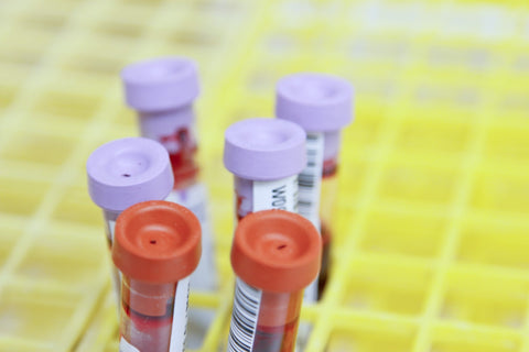
Collection of blood from the patient is a key step affecting the quality of the PRF produced. Care should be taken in choosing the tube for collecting the blood sample, as additive coatings (most commonly silica and/or silicone) on plastic centrifuge tubes can adversely affect the quality of the PRF.
Detailed studies of PRF clots prepared in different types of tubes have revealed five major drawbacks of silica- and/or silicone-coated tubes [4]:
- Silica-coated plastic tubes yield PRF clots that are generally more than 2× smaller than those from glass tubes (using the same volume of blood);
- Silica coatings are shed into PRF clots at “quite significant levels”, meaning that silica could end up in the patient’s body;
- Silica coatings cause cell death and reduce cell proliferation, impairing the wound-healing properties of the PRF;
- Clot formation occurs more slowly in plastic coated tubes than in glass tubes;
- PRF prepared in chemical-coated plastic tubes is reported to increase signs of inflammation when applied to a wound.
Therefore, if possible, whole blood from the patient should be collected into chemical-free glass tubes or glass-coated plastic tubes to ensure that the PRF produced is of a high quality.
Centrifugation
Centrifugation must be performed as soon as possible after collection to separate and concentrate the components of the blood before it clots.
Centrifugation is performed at room temperature to separate the patient’s whole blood into three main layers based on the density of the components. These are as follows, from top to bottom (least dense to most dense):
- Platelet-poor plasma (PPP) liquid layer;
- PRF clot, enriched in leukocytes, platelets, and growth factors;
- Red blood cell layer (also termed the red corpuscules base).
In addition, a buffy coat layer containing a high proportion of leukocytes is often found at the interface between the PRF clot layer and the red blood cell layer.
Horizontal vs. fixed-angle centrifuges
Selecting the correct centrifuge is critical to optimizing the quality of the PRF produced. So which type of centrifuge—horizontal or fixed-angle—is better for the job?
The answer from the scientific literature is clear, with studies conclusively showing that horizontal centrifuges are superior to fixed-angle centrifuges [5,6].
Horizontal centrifuges offer four main advantages over fixed-angle centrifuges.
Firstly, they separate the blood layers evenly, perpendicular to the tube, rather than at an angle—this facilitates the recovery of the PRF clot layer from the tube.
Secondly, the centrifugal force is applied through the blood sample, reducing trauma to cells as they move through the sample based on their relative densities. In a fixed-angle centrifuge, the cells are compressed against the side of the tube by the centrifugal force. This can lead to undesired shear stress to the cells or the aggregation of red blood cells, leukocytes, and platelets.
Thirdly, as the centrifugal force is applied along the axis of the tube, this increases the difference in the relative centrifugal force between the top and the bottom of the tube, leading to improved separation between the layers.
Finally, the count and concentration of platelets and leukocytes is increased by up to 3.5× by horizontal centrifugation relative to fixed-angle centrifugation.
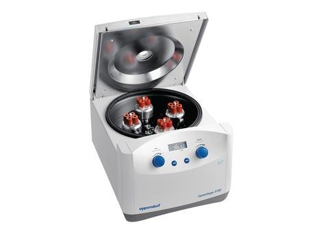
As PRF must be prepared from whole blood as quickly as possible, a benchtop centrifuge provides a convenient solution for rapidly centrifuging small amounts of blood from the patient. The Eppendorf 5702 centrifuge (above) has been found to produce an excellent separation of blood components and quality of PRF in a number of studies. [5,6,7]
Centrifugal force and duration
Selecting the appropriate force and duration of centrifugation is essential to obtaining a good distribution of wound-healing components in the PRF clot. In addition, depending on the centrifugation protocol used, either solid (L-PRF or A-PRF) or liquid (i-PRF) preparations of PRF can be produced.
In their study in 2020, Miron et al. systematically investigated the optimal centrifugal force and duration for preparing solid PRF (L-PRF or A-PRF) or liquid PRF (i-PRF) using the Eppendorf 5702 centrifuge [7]. The researchers compared 24 protocols involving six possible centrifugal forces (ranging from 100–1,200 g) and four possible durations (ranging from 3–12 min).
Their results showed that centrifuging at 400 g for more than 8 min led to an accumulation of leukocytes in the buffy coat layer, rather than throughout the upper PRF clot layers. At the other end of the spectrum, centrifugation protocols using 200 g or lower were unable to effectively concentrate platelets or leukocytes.
Solid PRF (L-PRF or A-PRF): After comparing all 24 protocols, they concluded that centrifugation at 700 g for 8 min is optimal for preparing solid PRF. This protocol produced PRF clots with the highest levels of platelets and leukocytes, which were evenly distributed throughout the clot.
Liquid PRF (i-PRF): Typically, i-PRF is produced using very low centrifugation speeds, such as 60 g, for 3–4 min. However, as shown in this and other studies [7,8,9], centrifugation at these low speeds is inadequate for concentrating platelets or leukocytes in the upper plasma layer. Rather, centrifugation at higher speeds of 200 g for 5 min [7] or 400 g for 3 min [9] have proven effective in maximizing platelet and leukocyte concentrations in a small volume of injectable plasma.
Conclusion
PRF offers significant advantages over PRP in terms of wound healing, thanks to the preservation of anticoagulants and the slow release of growth factors from the fibrin matrix. However, to optimize the benefits of the treatment and minimize any adverse effects on your patients, it is crucial to understand the best methods for preparing PRF.
Based on the up-to-date scientific evidence and practical recommendations in this article, we hope that your lab or clinic will be able to improve the quality of PRF it produces, supporting the speedy recovery and healing of your patients.
References
- Dohan DM, Choukroun J, Diss A, et al. Platelet-rich fibrin (PRF): A second-generation platelet concentrate. Part I: Technological concepts and evolution. Oral Surgery, Oral Medicine, Oral Pathology, Oral Radiology, and Endodontology. 2006;101(3):e37-e44. CrossRef Full Text
- Dohan Ehrenfest DM, de Peppo GM, Doglioli P, Sammartino G. Slow release of growth factors and thrombospondin-1 in Choukroun’s platelet-rich fibrin (PRF): a gold standard to achieve for all surgical platelet concentrates technologies. Growth Factors. 2009;27(1):63-69. CrossRef Full Text
- Ghanaati S, Booms P, Orlowska A, et al. Advanced Platelet-Rich Fibrin: A New Concept for Cell-Based Tissue Engineering by Means of Inflammatory Cells. Journal of Oral Implantology. 2014;40(6):679-689. CrossRef Full Text
- Miron RJ, Kawase T, Dham A, Zhang Y, Fujioka-Kobayashi M, Sculean A. A technical note on contamination from PRF tubes containing silica and silicone. BMC Oral Health. 2021;21(1):135. CrossRef Full Text
- Fujioka-Kobayashi M, Kono M, Katagiri H, et al. Histological comparison of Platelet rich fibrin clots prepared by fixed-angle versus horizontal centrifugation. Platelets. 2021;32(3):413-419. CrossRef Full Text
- Miron RJ, Chai J, Zheng S, Feng M, Sculean A, Zhang Y. A novel method for evaluating and quantifying cell types in platelet rich fibrin and an introduction to horizontal centrifugation. Journal of Biomedical Materials Research Part A. 2019;107(10):2257-2271. CrossRef Full Text
- Miron RJ, Chai J, Fujioka-Kobayashi M, Sculean A, Zhang Y. Evaluation of 24 protocols for the production of platelet-rich fibrin. BMC Oral Health. 2020;20(1):310. CrossRef Full Text
- Miron RJ, Fujioka-Kobayashi M, Hernandez M, et al. Injectable platelet rich fibrin (i-PRF): opportunities in regenerative dentistry? Clin Oral Invest. 2017;21(8):2619-2627. CrossRef Full Text
- Varela HA, Souza JCM, Nascimento RM, et al. Injectable platelet rich fibrin: cell content, morphological, and protein characterization. Clin Oral Invest. 2019;23(3):1309-1318. CrossRef Full Text