
What is a Percoll gradient?
Percoll is a silica colloid used to create density gradients. Percoll gradients are utilized in multiple life sciences fields for a variety of techniques and downstream applications. Their use in the separation and purification of immune cells from tissue and blood samples is perhaps the most widespread use, having been utilized for this purpose for over 40 years. Percoll gradients can be used to isolate specific cells, subcellular particles, and viral particles via centrifugation based on their individual densities.
Factors affecting gradient formation
Osmolality
To prevent cell swelling or shrinking, Percoll must be diluted in saline or cell culture medium, as appropriate, to generate an isotonic gradient with the physiological conditions of cells. The buoyant density of cells changes with the gradient osmolality - the concentration of all particles within the solution. Thus, it’s critical to ensure consistent osmolality across experiments and samples.
Diluent
For mammalian cells, saline or cell culture medium are appropriate diluents. However, subcellular components and viruses may aggregate in saline. For these purposes, Percoll can alternatively be diluted in sucrose. When hydrated, Percoll particles are smaller in the presence of 0.15 M NaCl than in 0.25 M sucrose and the sedimentation rate of the Percoll particles in saline is faster due to the lower viscosity.
Equipment
The centrifuge, rotor, and centrifuge tubes used will all influence the gradient shape and range of densities formed during centrifugation. Both time and g force determine gradient shape. Percoll diluted in 0.25 M sucrose will require greater g force to properly form gradients compared to that suspended in 0.15 M NaCl. The centrifuge must be able to reach at least 25,000 x g to facilitate Percoll gradient formation in sucrose.
For most applications, swinging bucket rotors are not recommended for generating Percoll gradients due to variations in force; a fixed-angle rotor is preferred. The rotor angle will greatly impact the shape and size of the gradient. Gradients generated in a near-vertical position will be shorter and will sediment more quickly than those at a more sloped angle.
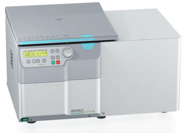
The HERMLE Z36-HK Super Speed Centrifuge with one of several high-speed fixed angle rotor options that fit tubes from 1.5 mL to 50 mL are suitable for a wide range of Percoll gradient applications.
Preparing Percoll Gradients
Gradients can be pre-formed before the isopycnic separation of cells at low speeds. However, viruses and subcellular particles generally will not efficiently separate on pre-formed gradients. In this case, in situ gradient preparation is needed where the sample is diluted in the Percoll solution prior to centrifugation; particles are separated simultaneously to gradient formation. It is generally a good idea when generating a Percoll gradient to simultaneously create an additional tube that will separate Density Marker Beads to serve as a method for assessing the gradient and determining density distributions.
Protocols will vary based on the tissue, target cell type, species, cell number, and application. Method-focused repositories and journals such as STAR protocols, BioProtocols, JoVE, and Cold Spring Harbor Protocols are great sources of protocols for many Percoll gradient applications.
Preparing a pre-formed gradient for cellular isolation
Cells can be isolated using pre-formed gradients or by directly suspending cell preparations in Percoll using a single-step protocol. A general protocol for separating cells on a pre-formed gradient is outlined below.
- Prepare a stock of 100% Percoll suspended in one 10th volume 10x PBS with final concentration of 10mM HEPES Buffer.
- Create the four-concentration gradient by diluting 100% Percoll stock in PBS. Load the highest concentration (50%) layer first, then carefully add the 40% and then 30% layers on top. For a 15-mL conical tube, each layer should be approximately 2-3 mL.
- Suspend the cells in 20% Percoll and immediately add to the conical tube as the top layer and proceed to centrifugation (500xg for 10 minutes). Note: protocols for the time and speed of centrifugation vary dramatically and will be affected by the cellular population you are isolating and the concentrations of Percoll present in your gradient.
In situ gradient preparation for viral isolation
Characteristics of the virus will impact the protocol required. One example of a protocol for virus isolation using a Percoll gradient is outlined below.
- Prepare a Percoll solution in a final concentration of 250mM sucrose.
- Suspend virus in Percoll solution. Note: a control tube with density marker beads should be used to calibrate Percoll gradient density.
- Centrifuge for 50,000xg for 25 min and then 100,000xg for 25 min. Remember: the gradient will continue to sediment during centrifugation.